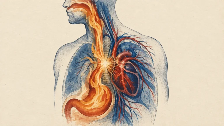
Your annual physical examination provides the ultimate opportunity to protect your independence and detect silent health risks before they cause lasting harm. Bone loss accelerates quietly as you age, often weakening your skeletal framework without producing a single ache or warning sign until a sudden fracture shatters your mobility. Dual-energy X-ray absorptiometry scans measure mineral density accurately and painlessly, allowing your physician to identify osteopenia or osteoporosis early and initiate targeted therapies. Requesting this painless imaging test during your upcoming wellness visit transforms routine preventive care into proactive protection against debilitating fractures, preserving your active lifestyle for decades to come.

Understanding Bone Density Testing: What You Need to Know
A dual-energy X-ray absorptiometry scan, commonly called a DXA scan, serves as the clinical benchmark for measuring bone mineral density. During this rapid, non-invasive examination, you lie comfortably on a padded table while a low-dose X-ray beam assesses your lumbar spine and hips. These central anatomical sites contain high proportions of trabecular bone, which exhibits metabolic changes earlier than cortical bone. The imaging system compares the attenuation of two distinct X-ray energies to calculate your precise mineral mass per square centimeter of bone tissue.
Physicians interpret your scan using diagnostic T-scores established by the World Health Organization. A T-score of -1.0 or higher reflects normal bone density; a score between -1.0 and -2.5 indicates osteopenia, or low bone mass; and a score of -2.5 or lower confirms osteoporosis. Surveillance data from the Centers for Disease Control and Prevention indicates that 12.6 percent of Americans aged fifty and older have osteoporosis, while 43.1 percent live with osteopenia. Knowing your score enables targeted interventions before structural failure happens.
Key Considerations: Why Bone Health Changes Dramatically with Age
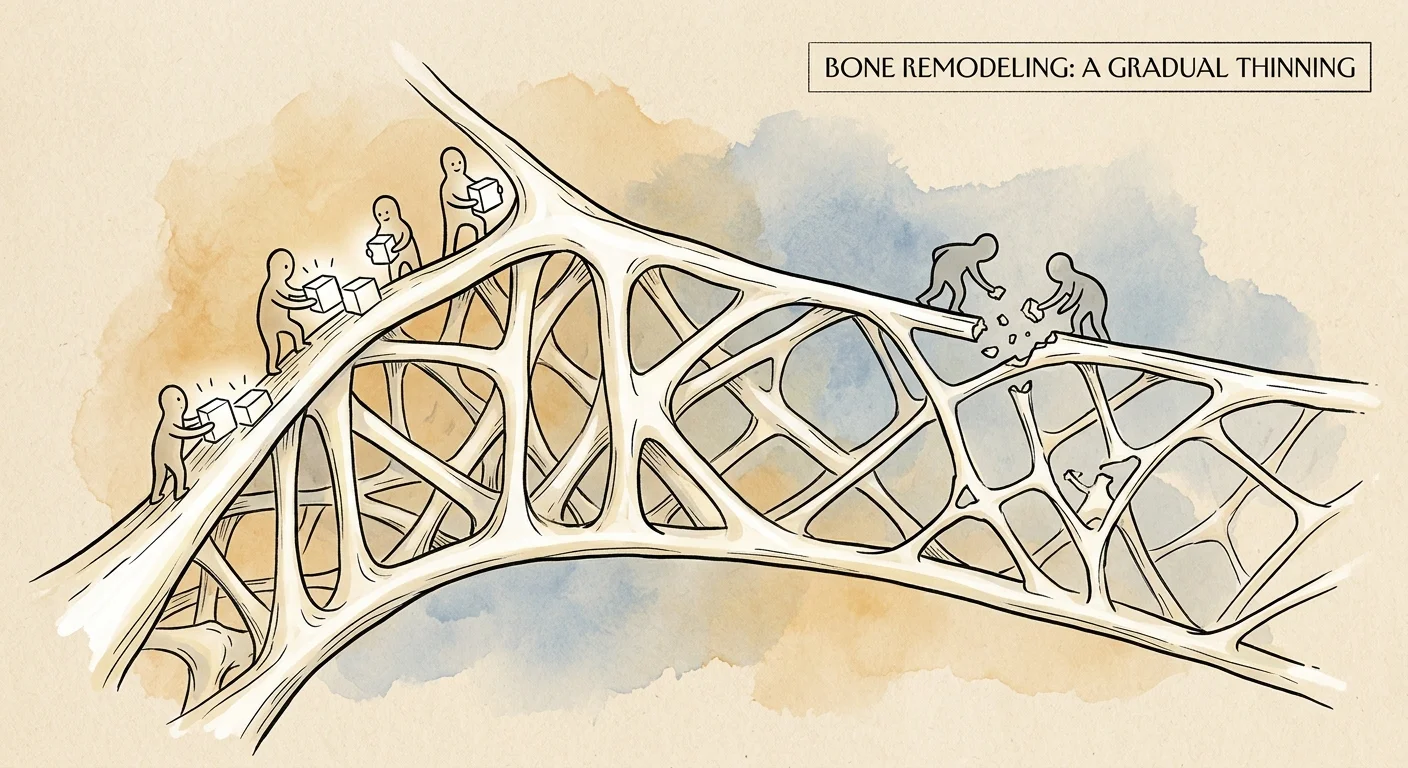
Your skeleton constantly renews itself through continuous remodeling, maintaining equilibrium between osteoclasts, which break down aged bone, and osteoblasts, which synthesize new bone matrix. After you achieve peak bone mass around age thirty, this delicate balance gradually tilts. Osteoclasts dismantle bone faster than osteoblasts can replace it, creating microscopic porosity within your skeleton. As the years advance, this structural thinning weakens bone architecture and substantially elevates fracture risks during routine daily activities.
Aging introduces several biological shifts that accelerate mineral depletion. Postmenopausal estrogen reduction removes a primary protective signal against bone resorption, while older men experience similar demineralization as testosterone levels drop. Simultaneously, age-related declines in stomach acid impair calcium absorption, and aging skin synthesizes less vitamin D. Coupled with muscle loss (sarcopenia), which reduces beneficial mechanical loading, fragile bones make older adults vulnerable to devastating hip and spinal fractures that threaten independent living.
8 Critical Signs Your Annual Physical Should Include a Bone Scan
Because osteoporosis progresses silently without early symptoms, recognizing clinical triggers helps you advocate for timely diagnostic testing. If any of these eight signs apply to you, request a bone scan during your physical examination.
Sign 1: You Have Reached the Standard Age Threshold for Routine Screening
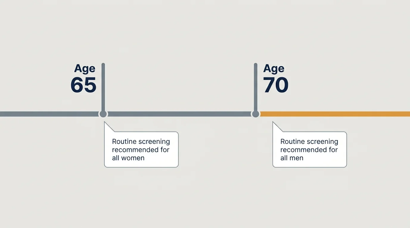
Reaching specific chronological milestones represents the most direct clinical trigger for bone screening. Guidelines from the U.S. Preventive Services Task Force and the Bone Health & Osteoporosis Foundation recommend routine DXA screening for all women aged 65 and older. For men, routine screening begins at age 70, although men aged 50 to 69 warrant testing if clinical risk factors exist. Age-related bone demineralization occurs predictably, making universal screening essential even if you feel vigorous.
Sign 2: You Notice Unexplained Height Loss or Postural Changes
Unexplained height reduction frequently signals silent spinal compression. Clinical guidelines recommend a bone density scan if you lose 1.5 inches (4 cm) or more compared to your young-adult height, or 0.8 inches (2 cm) between annual checkups. As vertebrae lose mineral density, ordinary daily weight bearing can cause painless wedge fractures that collapse anterior vertebral bodies, producing gradual height loss and a rounded thoracic stoop.
Sign 3: You Suffered a Fragility Fracture After Age 50
Breaking a bone after age fifty from minimal physical trauma demands immediate bone density evaluation. Clinicians define a fragility fracture as a break resulting from a fall from standing height or less, or from routine daily motions like coughing or lifting groceries. Healthy bones easily withstand minor trauma; breaking a wrist, hip, or rib during a slip reveals compromised bone strength and doubles your subsequent fracture risk.
Sign 4: You Rely on Long-Term Corticosteroid Medications
Taking oral corticosteroids like prednisone at doses of 5 milligrams or more daily for three or more consecutive months triggers an immediate recommendation for a baseline DXA scan. Glucocorticoids suppress osteoblast activity, accelerate osteoclast survival, and reduce intestinal calcium absorption. This medication-induced bone resorption causes rapid skeletal thinning, particularly in your spine and femoral neck, during the earliest months of therapeutic use.
Sign 5: You Experienced Early Menopause or Low Sex Hormone Levels
Sex hormones provide vital protection against rapid bone resorption. Undergoing natural or surgical menopause before age 45 deprives women of protective estrogen years ahead of schedule. Similarly, men with hypogonadism or depleted testosterone experience accelerated bone demineralization, because testosterone converts to estrogen to sustain male skeletal strength. If you have a history of premature ovarian failure or low hormone levels, request earlier screening.
Sign 6: Your Body Mass Index or Body Weight Is Exceptionally Low
Maintaining a very light frame increases vulnerability to bone loss. Adults with a body mass index below 19 kg/m² or a body weight under 127 pounds (57.6 kg) face significantly elevated fracture rates. Slender bodies provide less mechanical loading to stimulate bone remodeling during daily movement. Furthermore, individuals with low adipose tissue produce less peripheral estrogen, leaving fewer biological defenses against age-related mineral loss.
Sign 7: You Have a Direct Family History of Hip Fractures or Osteoporosis
Genetics heavily influence your skeletal geometry, cortical density, and peak bone mass. Having a biological parent who suffered a hip fracture or a major osteoporotic break significantly elevates your fracture probability, independent of your measured bone density. When direct family history indicates fragility fractures, clinical guidelines recommend screening before age 65 for women and before age 70 for men to establish a preventive baseline.
Sign 8: You Manage Chronic Illnesses or Take Bone-Depleting Prescriptions
Chronic inflammatory and metabolic disorders degrade bone quality over time. Conditions like rheumatoid arthritis, celiac disease, inflammatory bowel disease, primary hyperparathyroidism, and type 2 diabetes impair normal mineral balance. Concurrently, long-term prescription therapies—including aromatase inhibitors for breast cancer, androgen deprivation therapy for prostate cancer, selective serotonin reuptake inhibitors, and daily proton pump inhibitors for acid reflux—accelerate bone thinning. If you manage these factors, prioritize a bone evaluation.
Weighing the Benefits and Potential Risks of Bone Density Scans
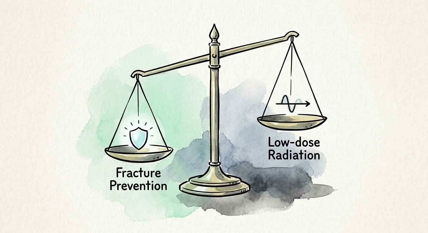
Evaluating the benefits and minor considerations of bone density scanning confirms its outstanding value in preventive geriatric medicine. The primary advantage of a DXA scan is early, accurate risk identification. Pinpointing low bone mass during the osteopenic stage enables you to implement dietary adjustments, targeted strength training, and medical therapies before catastrophic fractures occur. For those diagnosed with osteoporosis, baseline scans provide the objective metrics needed to monitor treatment efficacy over time.
The physical risks associated with bone density scans are negligible. A standard DXA scan emits an extraordinarily small radiation dose—roughly 1 to 10 microsieverts—which is less than a standard chest radiograph and comparable to the cosmic radiation absorbed during a cross-country flight. The main clinical consideration involves potential diagnostic artifacts: severe spinal osteoarthritis or aortic calcifications can falsely elevate lumbar readings. An experienced clinician or radiologist easily identifies these artifacts to ensure accurate diagnostic interpretation.
Medical Consensus: What Health Authorities Recommend
Major health organizations worldwide maintain uniform guidelines for bone density screening and fracture prevention. Consensus statements from the National Institutes of Health, the Endocrine Society, and the Mayo Clinic stress integrating bone scans into routine primary care for older adults. These organizations advocate using comprehensive evaluation tools, such as the Fracture Risk Assessment Tool (FRAX), which combines your DXA T-score with personal clinical risk factors to calculate your ten-year probability of suffering a major osteoporotic fracture.
When clinical risk models indicate a ten-year major fracture risk exceeding 20 percent, or a hip fracture probability surpassing 3 percent, professional guidelines recommend initiating prescription therapy. Health authorities emphasize that bone screening must guide a holistic prevention plan encompassing fall reduction strategies, nutritional optimization, and structured physical exercise to ensure continued physical autonomy.

Practical Steps and Actionable Advice for Your Wellness Visit
Taking proactive initiative during your annual physical ensures your bone health receives proper evaluation. Prior to your appointment, compile a concise medical record that details any past minor fractures, parental hip fractures, and all prescription medications or over-the-counter supplements you take daily. Measure your height at home to compare against your official medical record. When discussing your health goals, ask your physician directly whether your medical history, age, or lifestyle factors warrant ordering a DXA scan.
Alongside clinical testing, implement immediate lifestyle measures to fortify your skeleton. Aim for 1,200 milligrams of daily dietary calcium if you are a woman over fifty or a man over seventy, combined with adequate vitamin D3 to enhance mineral absorption. Perform weight-bearing activities like brisk walking and stair climbing, coupled with progressive resistance exercises three days weekly to stimulate bone formation. Finally, audit your home environment to eliminate trip hazards by securing rugs, improving lighting, and installing bathroom grab bars.
Frequently Asked Questions About Bone Density Screening
Understanding the details of bone mineral density testing helps you prepare for your physical with total clarity. Here are practical answers to the most common questions seniors ask.
How often should you repeat a bone density scan?
If your initial scan shows normal bone density and your risk profile remains low, repeating the test every three to five years is standard. If your scan identifies osteopenia or osteoporosis, or if you begin bone-modifying medication, clinicians recommend follow-up scans every two years to assess bone stability and monitor treatment response.
What should you expect during a dual-energy X-ray absorptiometry scan?
A DXA scan is completely painless, non-invasive, and lasts only ten to fifteen minutes. You lie clothed on an open, padded table while an imaging arm passes overhead. Wear clothing without metal fasteners or zippers, and avoid calcium supplements for twenty-four hours before testing to prevent imaging interference.
Does Medicare cover routine bone mineral density testing?
Medicare Part B covers diagnostic bone density screening once every twenty-four months—or more frequently when medically justified—for qualified beneficiaries. Eligible groups include estrogen-deficient women at clinical risk, individuals taking long-term oral corticosteroids, patients with primary hyperparathyroidism, and individuals taking FDA-approved osteoporosis medications to track therapeutic progress.
Why do older men need bone density scans?
Although osteoporosis is often perceived as a women’s health issue, one in five men over fifty will experience an osteoporotic fracture. Men who suffer hip fractures face higher rates of permanent disability and mortality within the first year than women. Age-related bone loss affects everyone, making testing critical for men aged 70 and older or those with clinical risks.
Can you rebuild bone density after receiving an osteoporosis diagnosis?
Yes; while you may not fully restore youthful peak bone mass, modern therapies significantly rebuild bone density and lower fracture risk. Anabolic medications stimulate fresh bone creation, while antiresorptive medications prevent further structural breakdown. When paired with progressive resistance training and optimized calcium and vitamin D intake, these interventions substantially strengthen your skeleton.
Taking Charge of Your Skeletal Health for Lifelong Vitality
Preserving your bone density protects your mobility, independence, and overall vitality as you age. A quick, painless DXA scan delivers essential data that transforms uncertainty into an effective, proactive health strategy. Discuss your bone health openly with your physician during your upcoming physical, evaluate your individual risk factors, and take decisive steps to keep your skeletal foundation strong for the road ahead.
This article is intended for informational and educational purposes only and is not a substitute for professional medical advice, diagnosis, or treatment. Always consult with a qualified healthcare provider regarding any health concerns or before making any decisions related to your health or treatment.