
Noticing new dark patches on your skin can feel alarming, but identifying their root cause gives you the power to protect your health. Most age-related pigment changes are harmless, yet certain dark spots signal underlying medical conditions that need professional attention.
Skin discoloration affects millions of older adults, shifting from minor cosmetic annoyances to important clinical clues. You can easily manage or treat these pigmentary changes once you understand what triggers them.
This practical guide breaks down the ten primary reasons behind uneven skin tone. You will discover how aging influences cellular melanin, which warning signs warrant prompt evaluation, and how modern dermatology treats persistent discoloration safely.
Understanding the Basics of Skin Discoloration
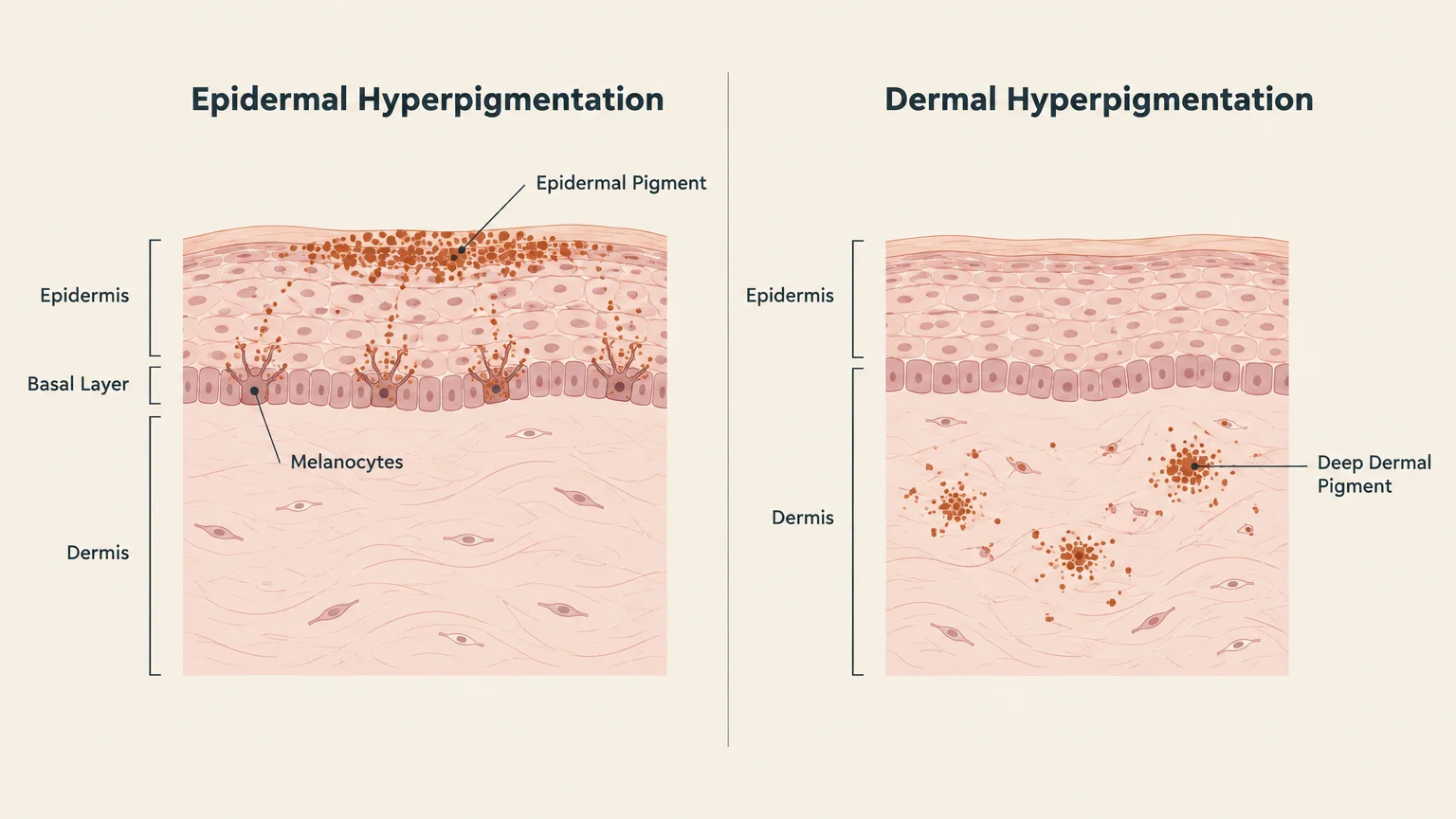
Your skin derives its unique color from melanin, a natural pigment produced by specialized cells called melanocytes. When these cells experience cellular stress, hormonal signals, or ultraviolet radiation, they often produce excess pigment.
Dermatologists refer to this localized darkening as hyperpigmentation. The process occurs when surplus melanin deposits unevenly across epidermal layers or sinks deeper into the dermis.
Understanding depth matters because surface-level epidermal pigment typically responds much faster to clinical interventions. In contrast, deeper dermal pigment often requires specialized therapies and patience to resolve completely.
Skin discoloration takes many distinct visual forms across different body regions. You might observe small, flat macules, broader diffuse patches, or textured, thickened plaques.
Differentiating harmless pigment accumulation from systemic illness or malignancy requires careful clinical observation. Evaluating changes in size, border regularity, and color depth provides vital clues about your cellular health.
Leading research institutions like the National Institutes of Health continue to study how cellular senescence accelerates localized pigment production. Knowing the biological mechanisms behind these marks empowers you to make informed decisions.
Melanocyte activity does not remain uniform across your lifespan. As decades pass, the total number of functioning melanocytes decreases, but surviving cells often cluster together and enlarge.
This irregular distribution explains why older adults frequently experience both localized dark spots and adjacent areas of pale, depigmented skin. Maintaining balanced skin health requires recognizing how these cellular dynamics shift over time.
10 Reasons You Might Be Getting Dark Patches on Your Skin
1. Chronic Sun Exposure and Solar Lentigines
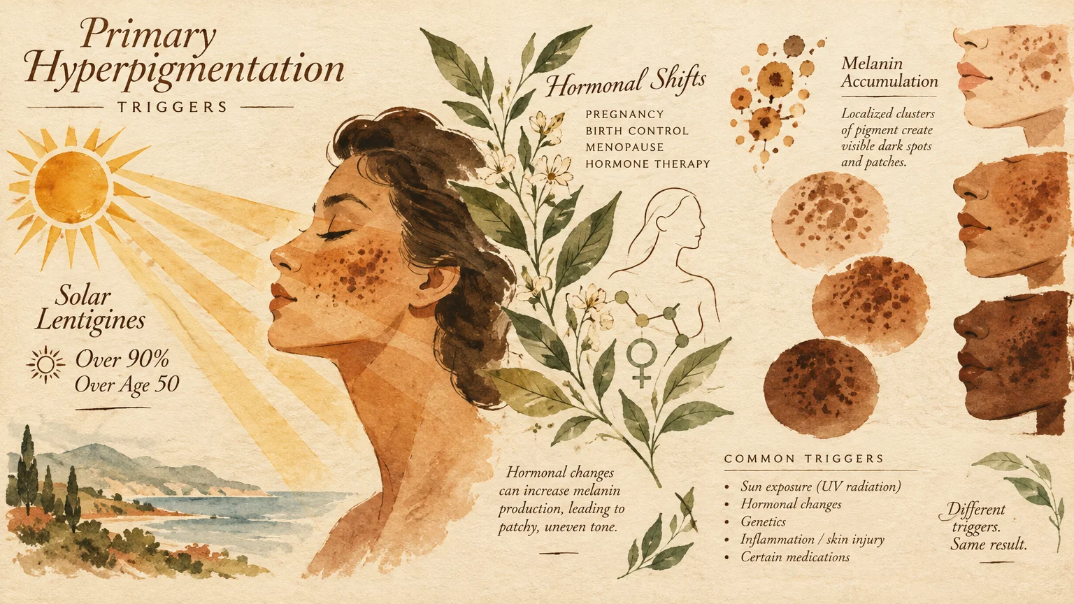
Decades of outdoor living inevitably leave their mark on your complexion. Solar lentigines, commonly known as sun spots or liver spots, represent concentrated clusters of melanin triggered by ultraviolet radiation.
These flat, tan, brown, or dark spots frequently appear on areas receiving direct sunlight. Your face, hands, forearms, and upper back represent prime real estate for these persistent lesions.
Clinical dermatology studies demonstrate that solar lentigines affect over 90% of fair-skinned individuals over the age of 50. In fact, this prevalence extends to up to 90% of people over age 60.
Ultraviolet rays damage the DNA within melanocytes, causing them to permanently overproduce pigment in concentrated clusters. Unlike childhood freckles, these age-related sun spots do not fade away during winter months.
While solar lentigines are entirely benign, their presence indicates significant cumulative sun exposure. Tracking them alongside your physician ensures they do not conceal more serious dermatological changes.
Fair skin types with less natural melanin baseline produce these spots more rapidly when exposed to sun. Consistent protective habits prevent existing spots from darkening further.
2. Hormonal Fluctuations and Melasma
Hormones exert a powerful influence over your melanocytes, frequently stimulating excess pigment production. Melasma presents as symmetrical, brownish-gray patches across the forehead, cheeks, upper lip, and bridge of the nose.
Global epidemiological data indicates that melasma affects roughly 1% of the general population overall. However, prevalence ranges from 9% to 50% in higher-risk populations, including individuals with darker phototypes.
Furthermore, up to 90% of melasma cases occur in women. Although frequently associated with pregnancy, mature women undergoing hormone replacement therapy often develop these distinct facial patches.
Estrogen and progesterone receptors on melanocytes amplify melanin synthesis when stimulated. When combined with incidental ultraviolet or visible light exposure, these hormonal triggers create stubborn, long-lasting discoloration.
Managing melasma requires dedicated, ongoing care because heat and light easily trigger sudden recurrences. Identifying hormonal factors with your medical team helps tailor an effective treatment plan.
Even short periods of sun exposure can undo months of diligent fading efforts. Gentle botanical lighteners and medical-grade mineral sunscreens offer relief without irritating vulnerable skin.
3. Post-Inflammatory Hyperpigmentation
Whenever your skin endures an injury, inflammatory cascade, or allergic flare, pigmentary changes can linger long after healing. Dermatologists identify this lingering discoloration as post-inflammatory hyperpigmentation.
Common culprits include eczema, contact dermatitis, shingles outbreaks, insect bites, or minor thermal burns. Even vigorous scratching or aggressive picking disrupts epidermal layers, prompting melanocytes to deposit surplus melanin.
This response occurs across all skin tones but tends to be more pronounced and persistent in deeper complexions. The resulting flat patches range in color from light brown to bluish-gray.
As skin matures, cellular turnover slows down, meaning these marks can linger for many months. Preventing mechanical trauma and treating inflammatory skin conditions promptly remains the most effective defense.
Topical soothing agents containing niacinamide or ceramides calm residual inflammation effectively. Avoiding harsh scrubs protects healing tissue from secondary trauma and pigment rebound.
4. Insulin Resistance and Acanthosis Nigricans
Sometimes, dark patches reveal critical information regarding your metabolic health and glucose processing. Acanthosis nigricans manifests as thick, velvety, hyperpigmented plaques found primarily in skin folds and creases.
You will most commonly notice these darkened bands along the back of the neck, armpits, and groin. Rather than feeling smooth, the affected skin develops a distinct, rippled, or velvet-like texture.
This condition links directly to hyperinsulinemia, where elevated circulating insulin stimulates epidermal keratinocytes and dermal fibroblasts. In national practice-based research, patients presenting with acanthosis nigricans are roughly twice as likely to have Type 2 diabetes compared to those without it.
Specifically, studies record a diabetes prevalence of 35.4% among patients with these patches versus 17.6% in unaffected controls. Furthermore, among women with Polycystic Ovary Syndrome, 60% to 80% display metabolic complications that include acanthosis nigricans.
Recognizing these velvety patches prompts vital laboratory screenings for prediabetes, metabolic syndrome, and endocrine imbalances. Addressing underlying insulin sensitivity through diet and medication often lightens the skin over time.
Scrubbing these dark bands with soap or exfoliants will not remove the discoloration and can cause painful irritation. Restoring metabolic balance remains the only reliable method to smooth and lighten affected folds.
5. Medication Side Effects and Drug-Induced Pigmentation
Many seniors take daily medications that inadvertently influence melanocyte activity or cause direct tissue deposition. Drug-induced hyperpigmentation accounts for a substantial percentage of unexpected skin discoloration in older adults.
Certain cardiovascular medications, such as amiodarone, can cause a slate-gray or bluish discoloration on sun-exposed skin. This occurs when drug metabolites accumulate within dermal macrophages following prolonged therapy.
Long-term use of antibiotics like minocycline, nonsteroidal anti-inflammatory drugs, and specific psychotropic medications can also stimulate pigment changes. Some antimalarial agents produce distinctive blue-black patches on the shins and oral palate.
Additionally, certain chemotherapy agents and targeted cancer therapies trigger localized or diffuse hyperpigmentation patterns. You should always review your complete medication list with a physician before stopping any prescribed treatment.
Drug-related pigmentation often stabilizes or slowly fades once your doctor substitutes the triggering pharmaceutical. Consistent broad-spectrum sun protection prevents these drug-induced deposits from darkening further under ultraviolet exposure.
6. Nutritional Deficiencies
Your body requires specific micronutrients to regulate cellular metabolism and maintain balanced skin tone. Severe or prolonged nutritional deficiencies can directly disrupt melanogenesis, producing patchy discoloration.
A deficiency in vitamin B12 remains a well-documented cause of localized and generalized hyperpigmentation. Adults over 60 frequently experience diminished gastric intrinsic factor, impairing dietary vitamin B12 absorption.
This deficiency often manifests as dark patches on the knuckles, palms, soles, and flexural folds. Fortunately, correcting your vitamin B12 levels through oral supplements or intramuscular injections typically resolves the discoloration.
Folate and iron deficiencies can also impair tissue oxygenation and cellular repair, causing an uneven, sallow complexion. Routine blood panels ordered by your primary care doctor quickly detect these reversible nutritional gaps.
Eating a nutrient-dense diet rich in leafy greens, lean proteins, and fortified foods supports skin repair. When malabsorption is present, working with a nutritionist helps identify therapeutic supplementation protocols.
7. Friction and Chronic Mechanical Irritation
Repeated physical pressure and friction against delicate skin stimulate protective thickening and pigment accumulation. Clinicians refer to this localized reaction as frictional melanosis or lichen simplex chronicus.
Tight-fitting clothing, rough elastic bands, heavy belts, and orthopedic braces frequently rub against vulnerable skin folds. Over time, your body responds to this ongoing mechanical irritation by generating excess keratin and melanin.
The inner thighs, underarms, and lower abdomen are especially prone to friction-induced darkening. This issue occurs more frequently in areas where skin rubs against skin during daily walking.
Switching to soft, breathable fabrics and applying protective barrier ointments significantly minimizes abrasive friction. Once you remove the mechanical stressor, the darkened skin gradually softens and fades.
Moisturizers enriched with colloidal oatmeal or dimethicone reduce friction while rebuilding the lipid barrier. Allowing irritated areas to heal without further rubbing accelerates natural skin lightening.
8. Adrenal and Endocrine System Disorders
Systemic endocrine disorders alter critical signaling hormones, leading to unexpected changes in pigment production. Addison’s disease, or primary adrenal insufficiency, represents a vital medical cause of widespread hyperpigmentation.
When your adrenal glands fail to produce sufficient cortisol, your pituitary gland compensates by releasing adrenocorticotropic hormone. Because this hormone shares structural similarities with melanocyte-stimulating hormone, it directly triggers systemic melanin overproduction.
Patients often notice hyperpigmentation in skin creases, pressure points, surgical scars, and inside the oral mucosa. Experiencing darkened gums alongside persistent fatigue and salt cravings warrants an immediate endocrine evaluation.
Thyroid disorders can also destabilize metabolic equilibrium and manifest through patchy facial or flexural discoloration. Balancing your hormone levels through targeted medical therapy remains the primary path to restoring healthy skin.
Endocrine-related skin changes often serve as an early clinical marker for internal hormone imbalances. Correcting underlying glandular health reverses widespread pigment surges safely and effectively.
9. Chronic Venous Insufficiency and Stasis Dermatitis
As veins in the lower legs age, their one-way valves may weaken and struggle against gravity. Chronic venous insufficiency causes blood to pool in your lower extremities, increasing vascular pressure significantly.
Under this elevated hydrostatic pressure, red blood cells escape delicate capillary walls and enter surrounding tissues. As these red blood cells break down, iron-rich hemosiderin pigment deposits permanently into the dermis.
This process produces mottled, reddish-brown, or rusty patches known as stasis dermatitis. You will typically see this discoloration developing around your ankles, shins, and calves.
Without intervention, the affected tissue can harden, itch intensely, and eventually break down into venous ulcers. Compression stockings, leg elevation, and vascular care alleviate pressure and prevent further pigment deposition.
Daily gentle moisturizing helps preserve fragile skin integrity across swollen lower legs. Vascular specialists can also perform minimally invasive procedures to restore healthy venous blood flow.
10. Precancerous Lesions and Skin Malignancies
While many dark patches are benign, differentiating harmless marks from skin cancer is critical for your longevity. As skin matures, the lifetime risk of developing cutaneous malignancies increases significantly.
Lentigo maligna is an early, non-invasive form of melanoma that frequently presents as a slowly enlarging dark patch. It appears most commonly on sun-damaged areas such as the face, neck, and ears.
Unlike benign sun spots, these lesions display irregular borders, uneven coloring, and asymmetrical growth patterns over time. Prompt surgical excision ensures complete removal before cancerous cells invade deeper dermal layers.
Seborrheic keratoses also appear frequently in older adults as waxy, dark, elevated plaques that look stuck onto the skin. While completely harmless, their dark appearance easily mimics melanoma, making a professional evaluation indispensable.
Actinic keratoses represent another precancerous growth that can occasionally present with pigmented, rough surfaces. Catching these lesions early through routine dermatologic mapping protects your long-term wellness.

Key Considerations for Seniors
Aging skin undergoes structural and physiological transformations that alter how pigment behaves and responds to treatment. The epidermal layer naturally thins over time, making underlying pigment and blood vessels far more visible.
At the same time, cellular turnover slows down substantially as you cross into your sixties and seventies. While younger skin sheds pigmented cells within weeks, mature skin requires double the time to regenerate.
Consequently, stubborn dark spots linger much longer after minor abrasions, chemical peels, or inflammatory episodes. Patience and gentle therapeutic strategies are essential to avoid worsening existing hyperpigmentation.
Seniors also face heightened risks of polypharmacy, where multiple daily prescriptions interact within the body. Drug interactions can increase photosensitivity, amplifying the darkening effect of incidental sun exposure.
Furthermore, weakened capillary fragility in mature skin leads to frequent micro-bruising and hemosiderin staining. Recognizing how your age impacts skin resilience helps you choose safer, more effective treatments.
Barrier repair becomes paramount when managing discoloration in older adults. Emollient creams containing ceramides and fatty acids fortify mature skin against environmental irritants.
Avoid harsh, drying alcohol-based toners or abrasive physical scrubs that scratch fragile epidermal barriers. Nourishing mature skin gently preserves its protective mantle while lightening stubborn marks.
Benefits and Potential Risks of Treating Dark Patches
Seeking treatment for dark patches can restore your skin tone and provide tremendous peace of mind. However, navigating the landscape of skin-lightening products requires extreme caution and clinical guidance.
Under the CARES Act, enacted in March 2020 and enforced starting September 23, 2020, over-the-counter hydroquinone products are illegal to sell in the United States. The FDA reclassified OTC hydroquinone as an unapproved drug due to significant health concerns.
Unregulated hydroquinone carries serious risks, most notably exogenous ochronosis, which causes permanent bluish-black skin discoloration. Today, hydroquinone is legally available only through a doctor’s prescription, such as Tri-Luma or 503A compounding pharmacies.
Additionally, the FDA maintains active consumer safety alerts warning against unapproved, imported, or illegally marketed skin-lightening creams. Laboratory testing since 2019 has repeatedly identified dangerous levels of hydroquinone and toxic mercury in these products.
Absorbing topical mercury damages the nervous system, impairs kidney function, and poses severe systemic health threats. You must avoid unverified creams purchased online or from unregulated international distributors.
Legitimate dermatological therapies deliver substantial aesthetic improvements when administered by licensed medical practitioners. Clinically supervised options include topical retinoids, azelaic acid, tranexamic acid, and gentle laser treatments.
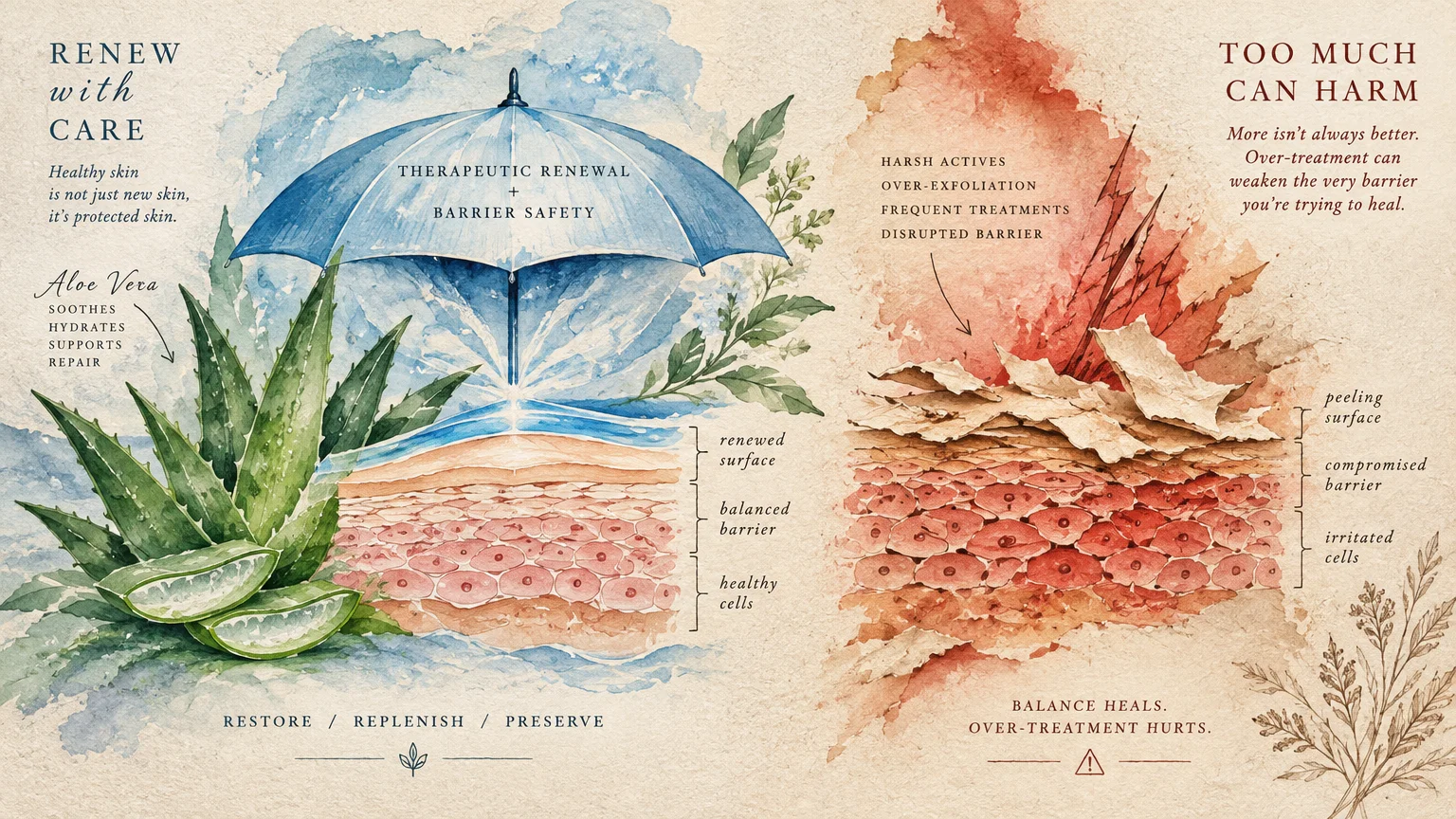
Nevertheless, aggressive chemical peels or inappropriate laser settings can induce severe post-inflammatory hyperpigmentation. A measured, medically guided strategy prevents unwanted complications while gently clearing persistent dark spots.
Professional oversight ensures that active ingredients brighten discoloration without triggering barrier breakdown or rebound darkening. Prioritizing safety over quick fixes protects both your appearance and your overall health.
What the Experts Say
Leading medical organizations emphasize that daily photoprotection forms the essential cornerstone of hyperpigmentation management. Preventing ultraviolet and visible light from reaching melanocytes stops the pigment cascade before it begins.
For preventing UV-induced dark spots and melasma flares, the American Academy of Dermatology recommends daily use of a broad-spectrum, water-resistant sunscreen with an SPF of 30 or higher. Consistency remains far more critical than relying on expensive corrective serums alone.
For melasma and post-inflammatory hyperpigmentation specifically, dermatologists recommend tinted sunscreens containing iron oxides. Iron oxides block visible light, which researchers now recognize as a potent trigger for pigment production in darker skin tones.
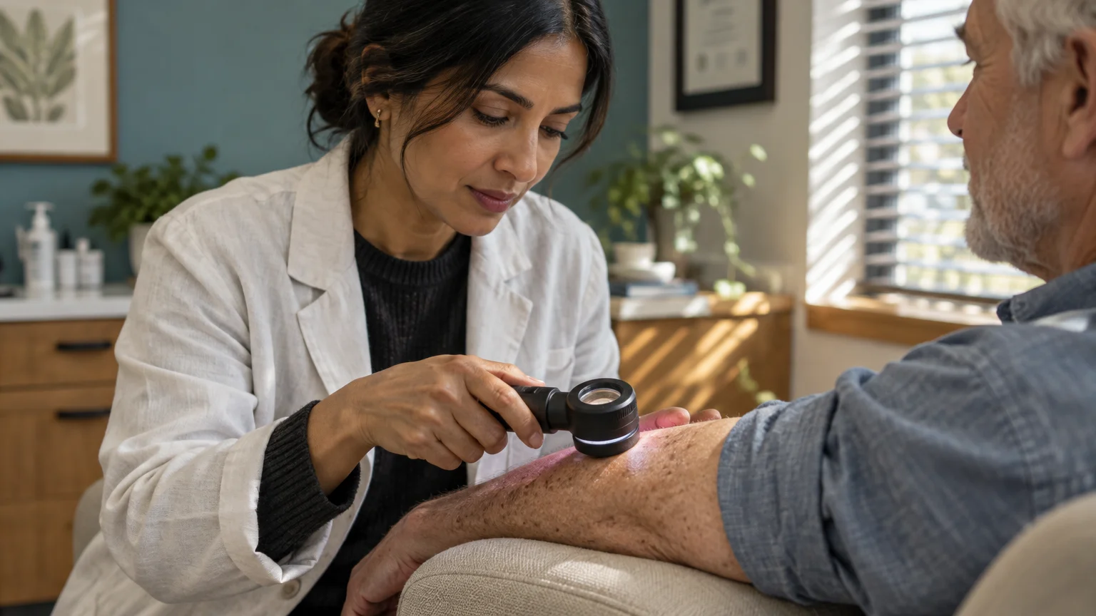
Specialists at the Mayo Clinic stress the importance of annual full-body skin examinations for adults over 60. Having a dermatologist examine your skin under dermoscopy distinguishes benign solar lentigines from early-stage melanomas accurately.
The Centers for Disease Control and Prevention reminds older adults that broad sun safety includes shade and protective clothing alongside sunscreen. Comprehensive sun defense dramatically slows cellular aging and minimizes hyperpigmentation.
Researchers at the World Health Organization also caution against untested cosmetic lightening agents worldwide. Global health bodies urge patients to rely solely on scientifically verified medical treatments.
Consensus across international dermatology societies confirms that gentle, sustained interventions yield the best outcomes. Quick-fix bleaching remedies often create severe rebound darkening that is exceedingly difficult to reverse.

Practical Steps and Actionable Advice
Taking proactive control of your skin health does not require complicated routines or risky procedures. Adopting evidence-based habits protects your skin barrier while systematically reducing stubborn discoloration.
Apply a broad-spectrum sunscreen with an SPF of 30 or higher every single morning, regardless of cloud cover. Choose a tinted mineral formula with iron oxides to shield your skin from both ultraviolet and high-energy visible light.
Incorporate physical sun protection into your daily routine when gardening, golfing, or walking outdoors. Wide-brimmed hats, ultraviolet-protection factor clothing, and polarized sunglasses provide continuous defense without requiring reapplication.
Conduct a thorough self-examination of your skin once each month in a brightly lit room. Use a full-length mirror and a hand mirror to inspect hard-to-see areas like your back, scalp, and buttocks.
Utilize the ABCDE framework to evaluate suspicious spots: look for Asymmetry, Border irregularity, Color variation, Diameter over six millimeters, and Evolving shape. Schedule a professional evaluation immediately if any spot changes size, shape, or shade.
Gather your current prescription and over-the-counter medications to review with your primary care physician. Ask directly whether any of your routine pharmaceuticals list photosensitivity or cutaneous hyperpigmentation as potential side effects.
If you notice velvety, darkened skin around your neck or armpits, request comprehensive metabolic laboratory testing. Fasting glucose, hemoglobin A1c, and fasting insulin tests clarify whether insulin resistance requires dietary or pharmaceutical intervention.
Avoid the temptation to buy unregulated lightening creams online or from unverified international vendors. Always obtain skin-brightening treatments through a board-certified dermatologist or licensed healthcare provider.
Keep a daily skin journal if you experience recurring patches of irritation or discoloration. Noting cosmetic products, clothing fabrics, and outdoor activities helps your physician pinpoint subtle triggers quickly.
Frequently Asked Questions
Can dark spots on my skin turn into melanoma?
Benign sun spots and seborrheic keratoses do not mutate into melanoma over time. However, early melanomas frequently resemble common age spots, making professional dermoscopic exams critical for accurate diagnosis.
Never assume that a flat brown spot is merely a sun spot without having it evaluated. If an existing spot begins expanding, darkening, or bleeding, schedule an appointment immediately.
Are age spots permanent, or can you fade them?
Age spots are permanent unless actively treated with targeted dermatological therapies. Prescription topical retinoids, clinical chemical peels, cryotherapy, and laser treatments can safely fade or eliminate them.
Even after successful removal, new spots will develop if you expose your unprotected skin to ultraviolet rays. Consistent daily sunscreen application prevents faded age spots from returning.
Why are over-the-counter hydroquinone creams no longer sold in stores?
The FDA removed OTC hydroquinone from store shelves under the CARES Act due to safety concerns. Prolonged unregulated use poses risks of permanent ochronosis discoloration and toxic contamination in imported formulations.
Hydroquinone remains available legally only through a physician’s prescription, ensuring your treatment is carefully monitored. This regulatory oversight protects consumers from counterfeit or contaminated products.
How does diabetes cause dark, velvety skin patches?
Elevated circulating insulin levels stimulate rapid multiplication of skin cells and fibroblasts. This localized cell growth produces the characteristic velvety, hyperpigmented plaques known clinically as acanthosis nigricans.
Improving blood glucose control through medication and lifestyle changes lowers circulating insulin levels. Over several months, this metabolic stabilization allows darkened skin folds to soften and lighten significantly.
Does changing my diet help clear dark patches on the skin?
Dietary adjustments directly improve dark patches caused by metabolic conditions like acanthosis nigricans. Reducing refined carbohydrates and balancing blood glucose lowers insulin levels, which gradually clears the velvety discoloration.
Similarly, resolving verified vitamin B12 or iron deficiencies through nutrient-rich foods or supplements reverses nutritional hyperpigmentation. However, diet alone cannot erase sun-induced lentigines without dedicated sun protection.
Moving Forward with Confidence and Clear Skin
Developing dark patches on your skin is a widespread, natural component of the aging journey. Recognizing whether these marks stem from sun exposure, hormonal shifts, metabolic changes, or medications empowers you to take charge.
Proactive sun defense remains your most formidable tool for preserving an even skin tone and shielding cellular DNA. Combining daily broad-spectrum tinted sunscreen with routine self-checks protects both your appearance and your overall vitality.
Never hesitate to bring new, shifting, or unusual dark spots to the attention of a board-certified dermatologist. Clear communication with your medical team ensures you receive safe, evidence-based treatments tailored to your unique skin.
This article is intended for informational and educational purposes only and is not a substitute for professional medical advice, diagnosis, or treatment. Always consult with a qualified healthcare provider regarding any health concerns or before making any decisions related to your health or treatment.